Intraoral Camera
Temecula, CA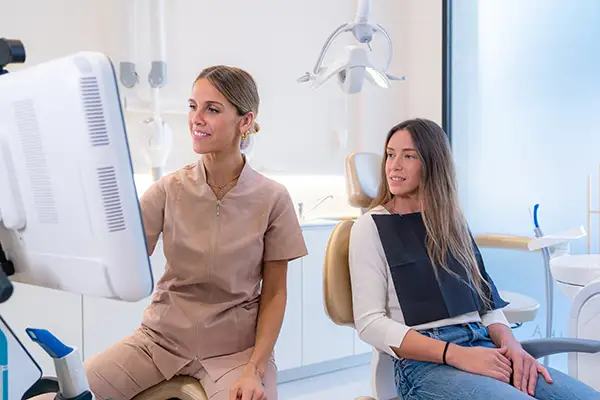
Most patients have seen an intraoral camera in action without necessarily knowing how the device behind that small wand actually works. At Picasso Dental Care, we rely on this dental technology every day, and patients often ask what is happening on the other end of that thin, pen-shaped instrument as it moves gently around their mouth. Understanding the mechanics behind intraoral cameras helps explain why the images they produce are so clear, and why that clarity translates directly into better dental care.
The Camera Itself
An intraoral camera is built around a tiny, high-resolution image sensor housed inside a slim, lightweight wand. The wand is shaped to glide comfortably along the surfaces of the teeth and gums, reaching areas that are difficult to view directly, even with a mirror and good lighting. The sensor at the tip captures a continuous stream of images as the wand moves, rather than a single static photograph, which allows our dental team to explore the entire mouth methodically without missing sections.
From Lens To Screen
Once the sensor captures an image, that data travels through a cable or wireless connection to a computer or monitor positioned within view of the dental chair. The transfer happens instantly, so what the camera sees appears on screen in real time. This live feed is what allows the dentist to narrate findings as they happen, pointing out specific areas of interest while the patient watches the same image at the same moment, rather than waiting for a description after the fact.
Lighting And Magnification
The inside of the mouth is a dim, tightly confined space, which is part of why intraoral cameras include their own built-in light source positioned directly at the tip of the wand. This integrated lighting eliminates shadows that might otherwise obscure small details. Combined with magnification that can enlarge a tooth surface many times its actual size, the camera reveals fine details such as hairline cracks, early decay, or subtle changes in gum tissue that would be nearly impossible to see with the naked eye alone, even under a typical overhead exam light.
Built For Comfort And Precision
Patient comfort plays a real role in how intraoral cameras are designed. The wand is intentionally lightweight and slim, often no larger around than a standard pen, so it can be guided around the mouth without triggering a gag reflex or causing discomfort. Many designs also include a disposable or sterilizable sheath that covers the wand for each patient, keeping the device clean and ready for use. The smooth, rounded tip is shaped to avoid catching on soft tissue, and the cable is flexible enough to allow our team to angle the camera precisely where it is needed.
Connecting To Digital Records
Once captured, images from the intraoral camera do not simply disappear after the appointment ends. They can be saved directly into a patient's digital chart, creating a visual record that sits alongside notes, x-rays, and treatment history. This means that if a small area of concern is being monitored over several visits, our team can pull up a previous image and compare it directly to a new one, looking for any change in size, color, or texture. That side-by-side comparison is far more precise than relying on memory or written notes alone.
Why The Technology Matters
None of these technical details matter much on their own. What matters is what they make possible during your visit. Because the images are captured instantly, displayed in real time, and stored for future reference, intraoral cameras allow our dentists to identify problems earlier, explain findings more clearly, and track changes in your oral health with a level of accuracy that was simply not available a generation ago. The technology itself is sophisticated, but the experience for the patient is straightforward: a brief, painless scan that produces images you can see and understand right alongside your dentist.
A Tool That Supports Better Conversations
Beyond diagnosis, the camera changes the nature of the conversation that happens in the exam chair. Instead of being told that a tooth has a small crack or that a filling is starting to wear, patients can see exactly what that looks like on screen. This shared view tends to make treatment recommendations easier to understand and easier to accept, because the evidence is visible rather than simply described. It also gives patients a chance to ask more specific questions, since they are looking at the same image as the dentist rather than relying solely on a verbal explanation.
How The Images Support Treatment Planning
The detail captured by an intraoral camera does more than confirm a diagnosis in the moment. When the dentist is mapping out a treatment plan, having a clear, magnified image of the affected area makes it easier to decide on the right approach, whether that means a conservative filling, a more involved restoration, or simply continued monitoring. Because the image can be reviewed closely after the exam, treatment decisions are based on precise visual information rather than a quick glance or a brief verbal description recalled from memory.
A Technology That Continues To Improve
Intraoral camera systems have evolved considerably since they were first introduced into general dentistry. Early versions produced grainy, low-resolution images that offered only a modest improvement over the naked eye. Today's cameras use high-definition sensors capable of capturing crisp detail, and many systems now include software that can adjust contrast, color balance, and focus automatically as the wand moves. This steady improvement in image quality means that the small details our team relies on for early detection are easier than ever to see clearly.
At Picasso Dental Care, we use intraoral camera technology because it lets us practice more precise, more transparent dentistry. If you would like to see how this technology works during your own exam, call us at (951) 383-2021 to schedule a visit. We are glad to walk you through what we see and why it matters for your smile.
|