3D Imaging
Temecula, CA
Jaw pain, clicking, and difficulty opening or closing the mouth comfortably are common reasons patients visit Picasso Dental Care. The temporomandibular joint, or TMJ, is a complex structure, and the source of discomfort is not always obvious from a clinical dental exam alone. Three-dimensional imaging allows our team to examine the joint itself, along with the surrounding bone, in detail that a standard X-ray simply cannot provide.
Why the TMJ Is Difficult to Evaluate
The temporomandibular joint connects the lower jaw to the skull and allows the wide range of motion needed for chewing, speaking, and yawning. Because it is a small joint made up of bone, cartilage, and a disc that moves with each jaw motion, evaluating it accurately requires more than a glance at a flat image. Traditional panoramic X-rays show the general shape of the jaw, but they offer limited detail about the joint surfaces themselves, the space between the bones, or subtle changes that may explain a patient's symptoms.
This is part of why TMJ disorders can be challenging to diagnose. Symptoms such as clicking, popping, or a sensation of the jaw catching can stem from several different causes, ranging from disc displacement to joint degeneration to muscular tension affecting jaw function. Without detailed imaging, it can be difficult to distinguish between these possibilities, which makes it harder to recommend the most effective treatment.
What 3D Imaging Shows
Cone beam computed tomography produces a detailed, three-dimensional view of the jaw joint and the bone structures surrounding it. Our team can examine the joint from multiple angles, measure the space within it, and assess the condyle, the rounded end of the lower jaw bone that fits into the joint socket.
This level of detail allows us to identify findings that directly inform treatment, including:
| • |
Changes in the shape or surface of the condyle that may indicate degenerative joint changes |
| • |
Asymmetry between the left and right joints, which can contribute to bite and jaw function issues
|
| • |
Narrowing of the joint space, which may relate to disc position or joint compression
|
| • |
Bone remodeling or erosion associated with long-term joint strain
|
| • |
The relationship between the jaw joint and surrounding structures, including the ear canal and skull base |
By examining these structures in three dimensions, our team gains a far clearer understanding of what may be contributing to your symptoms, rather than relying on guesswork based on a flat image or symptoms alone.
Connecting Imaging Findings to Treatment
Once a 3D scan has been reviewed, our team can use the findings to guide a treatment approach suited to the specific issue identified. For some patients, this may mean a conservative approach such as a custom nightguard or bite splint designed to reduce strain on the joint. For others, imaging may reveal a more significant structural issue that benefits from a different course of care, including referral to a specialist.
Having detailed imaging available also helps our team track how a patient's joint responds to treatment over time, giving both our team and the patient a clearer sense of progress rather than relying solely on self-reported comfort.
Distinguishing Joint Problems From Muscle-Related Pain
One of the most useful aspects of three-dimensional imaging is its ability to help separate true joint problems from pain that originates in the surrounding muscles. Many patients who experience jaw discomfort assume the joint itself is the source, when in some cases the muscles responsible for chewing and jaw movement are the primary contributor. Because the treatment approach for a structural joint issue can differ significantly from the approach for muscle-related tension, this distinction matters.
When a 3D scan shows a joint with a healthy condyle, normal joint space, and no signs of bone remodeling, that information helps our team look more closely at muscular or bite-related factors as the likely source. On the other hand, when imaging reveals clear structural changes, that finding shifts the conversation toward treatment options aimed directly at the joint itself. Either way, having objective imaging removes much of the uncertainty that can come with diagnosing TMJ-related symptoms based on a physical exam alone.
Supporting a Personalized Treatment Plan
No two patients experience TMJ symptoms in exactly the same way, and imaging helps ensure that treatment is tailored to the individual rather than applied as a general protocol. A patient with mild joint space narrowing and no significant bone changes may respond well to a simple bite splint, while a patient showing more advanced degenerative changes may need a more involved approach or referral to a TMJ specialist for further evaluation.
This individualized approach also helps set realistic expectations. When our team can show you what is happening within your joint, it becomes easier to understand why a particular treatment is being recommended and what signs would indicate the need for a different approach.
When TMJ Imaging Is Recommended
Not every patient with occasional jaw discomfort needs a 3D scan. Our team typically recommends this imaging when symptoms are persistent, when there is a noticeable change in jaw function, when clicking or popping is accompanied by pain, or when a patient has a history of trauma to the jaw. Imaging may also be helpful before starting orthodontic treatment in patients with existing jaw joint concerns, since shifting the bite can affect how the joint functions.
A Comfortable, Efficient Process
A cone beam scan of the jaw joint takes less than a minute to complete and involves significantly less radiation than a hospital CT scan. You remain in a comfortable, stationary position while the scanner captures images from every angle. Once the scan is complete, our team reviews the results and can walk you through what we are seeing, pointing out the specific areas relevant to your symptoms so you have a clear understanding of your own anatomy and what it means for your care.
Find Relief From Jaw Pain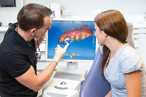
Living with ongoing jaw pain or joint dysfunction can affect everything from eating to speaking comfortably. At Picasso Dental Care, three-dimensional imaging gives our team the tools to identify what is truly happening within your jaw joint, supporting a treatment plan built around your specific anatomy rather than general assumptions. If you are experiencing jaw pain, clicking, or limited movement, call us today at (951) 383-2021 to schedule an evaluation. |